Trial published at NEJM
involve 180 pt Advanced Idiopathic Pulmonary Fibrosis (defined as a carbon monoxide diffusion capacity of < 35% of the predicted value)
sildenafil did not cause a significant difference in the proportion of patients with an improvement of 20% or more in the 6-minute walk distance at 12 weeks (the primary outcome). There were small differences favoring sildenafil in some secondary outcomes, including the degree of dyspnea and quality of life.
patients receiving sildenafil during period 1 had symptomatic benefit of arterial blood gas and carbon monoxide diffusion capacity, as compared with placebo-treated patients.
previously published data showing that sildenafil improved ventilation–perfusion matching
Implication:sildenafil was associated with symptomatic improvement may be of value to patients with advanced idiopathic pulmonary fibrosis.
Tuesday, May 25, 2010

Monday, May 24, 2010
Endovascular versus Open Repair of Abdominal Aortic Aneurysm The United Kingdom EVAR Trial Investigators
Cardiovascular versus Open Repair of Abdominal Aortic Aneurysm
The United Kingdom EVAR Trial Investigators
From 1999 through 2004 at 37 hospitals in the United Kingdom, involving 1252 patients with large abdominal aortic aneurysms (≥5.5 cm in diameter) to undergo either endovascular or open repair; 626 patients for each group.
Follow up 5-10 years.
30-day operative mortality was 1.8% in the endovascular-repair group and 4.3% in the open-repair group
benefit was lost by the end of the study, at least partially because of fatal endograft ruptures.
end of follow-up, there was no significant difference between the two groups in the rate of death from any cause.
CONCLUCION:patients who were considered to be suitable candidates for either endovascular repair or open repair of abdominal aortic aneurysm, the endovascular procedure was associated with a significantly lower operative mortality. However, no significant differences were seen in total mortality or aneurysm-related mortality in the long term. Endovascular repair was associated with increased rates of complications and reinterventions and was more costly
The United Kingdom EVAR Trial Investigators
From 1999 through 2004 at 37 hospitals in the United Kingdom, involving 1252 patients with large abdominal aortic aneurysms (≥5.5 cm in diameter) to undergo either endovascular or open repair; 626 patients for each group.
Follow up 5-10 years.
30-day operative mortality was 1.8% in the endovascular-repair group and 4.3% in the open-repair group
benefit was lost by the end of the study, at least partially because of fatal endograft ruptures.
end of follow-up, there was no significant difference between the two groups in the rate of death from any cause.
CONCLUCION:patients who were considered to be suitable candidates for either endovascular repair or open repair of abdominal aortic aneurysm, the endovascular procedure was associated with a significantly lower operative mortality. However, no significant differences were seen in total mortality or aneurysm-related mortality in the long term. Endovascular repair was associated with increased rates of complications and reinterventions and was more costly
Multiple Beau's Lines

Multiple Beau's Lines as picture above commonly seen in pt undergoing chemotherapy
as cytotoxic chemotherapeutic agents can induce the temporary arrest of proliferative function of the nail matrix.
it may suggest very severe illness too if only single beau's line is seen.
Multiple beaus line almost equal to malignancy with chemotherapy
bilateral ptosis
If you find bilateral ptosis in the patient examined
3 diagnosis must blink in your brain
1) GBS (guillain barre syndrome) if + complete opthalmoplegia then = Miller fischer
Check for ataxia and opthalmoplegia for miller fischer , areflexia for GBS
2) Myasternia grvis demonstrate eyelid fatigue, proximal myopathy fatigue and verbal fatigue(ask pt to count 1-50 continuously)
3) Dystrophy myotonica-demonstrate percussion myotonia at palmar and tongue.
or rare causes-oculopharyngeal dystrophy,
third nerve palsy caused by oculomotor nucleus lesion (rare), multiple sclerosis also may considered but normally presented with internuclear opthalmoplegia and optic atrophy.
Experience in exam:
a lady presented with weakness of bilateral lower limb. Please examine the lower limb.
surprisingly normal tone and power 5/5, reflex absent on jendrassic maneuver
request for gait , cerebellar sign , sensation and gait.
Examiner not keen.
Request to look at the face.
A lady with eyes closed, ask to open eyes demonstate eyes movement -complte opthalmoplegia
want to demonstrate ataxia. stopped by examiner
ask what else to consider -want to demonstrate fatigue of muscle-proceed no fatigue of muscle.
Final diagnosis- miller fischer
ask about feature of miller fischer?
give the famous three-opthalmoplegia, ataxia and areflexia.
3 diagnosis must blink in your brain
1) GBS (guillain barre syndrome) if + complete opthalmoplegia then = Miller fischer
Check for ataxia and opthalmoplegia for miller fischer , areflexia for GBS
2) Myasternia grvis demonstrate eyelid fatigue, proximal myopathy fatigue and verbal fatigue(ask pt to count 1-50 continuously)
3) Dystrophy myotonica-demonstrate percussion myotonia at palmar and tongue.
or rare causes-oculopharyngeal dystrophy,
third nerve palsy caused by oculomotor nucleus lesion (rare), multiple sclerosis also may considered but normally presented with internuclear opthalmoplegia and optic atrophy.
Experience in exam:
a lady presented with weakness of bilateral lower limb. Please examine the lower limb.
surprisingly normal tone and power 5/5, reflex absent on jendrassic maneuver
request for gait , cerebellar sign , sensation and gait.
Examiner not keen.
Request to look at the face.
A lady with eyes closed, ask to open eyes demonstate eyes movement -complte opthalmoplegia
want to demonstrate ataxia. stopped by examiner
ask what else to consider -want to demonstrate fatigue of muscle-proceed no fatigue of muscle.
Final diagnosis- miller fischer
ask about feature of miller fischer?
give the famous three-opthalmoplegia, ataxia and areflexia.
Thursday, May 20, 2010
Horner syndrome
Horner syndrome
confirm it is horner
"Everything is small"-ptosis,small pupil(miosis),anhydrosis and enolthalmos(occasionally)
proceed to horner protocol
check other cranial nerve to rule out Wallenberg(lateral medullary) syndrome, sensation 5th, facial nerve, gag reflex for 9th and 10th and cerebellar sign plus contralateral sensation .
If no, then look for radiation marking , wasting small muscle , dullness apical ocasionally engorged vessel (Superior vena cava obs) to suggest Pancoast tumour.
dissociated sensory loss and burn scar to suggest syringomyelia if gag reflex reduced than syringobulbia.
surgical scar at neck may suggeast injury to the T1 nerve.
Present the finding .
You should be safe for this station.
(P/S) may sure it is Horner before do all this.
confirm it is horner
"Everything is small"-ptosis,small pupil(miosis),anhydrosis and enolthalmos(occasionally)
proceed to horner protocol
check other cranial nerve to rule out Wallenberg(lateral medullary) syndrome, sensation 5th, facial nerve, gag reflex for 9th and 10th and cerebellar sign plus contralateral sensation .
If no, then look for radiation marking , wasting small muscle , dullness apical ocasionally engorged vessel (Superior vena cava obs) to suggest Pancoast tumour.
dissociated sensory loss and burn scar to suggest syringomyelia if gag reflex reduced than syringobulbia.
surgical scar at neck may suggeast injury to the T1 nerve.
Present the finding .
You should be safe for this station.
(P/S) may sure it is Horner before do all this.
Monday, May 17, 2010
Approach patient with unilateral ptosis
When You see patient with partial ptosis, One of the commonest patient found in neuro station. See whether it is unilateral or bilateral.
for unilateral
The second thing to do is look directly to pupil size,
If pupil size is small then you should enter Horner syndrome protocol which i will explain in my next explaination.
If pupil size is big then , it is third nerve palsy(a surgical third nerve palsy) most likely posterior communicating artery aneurysm.
If pupil size is normal then most likely (medical third nerve palsy) most likely diabetic or vasculitis cause and etc.
Third please demonstate the eye movement to see whether got other nerve involvement.etc 4th or 5th
If got 3rd ,4th and 5th, consider Graves disease(lid retraction )
cavernous sinus 3th 4th 6th and 5th may coexixst(demonstrate corneal reflex)
tolosa hunt (pain with no redness)
for unilateral
The second thing to do is look directly to pupil size,
If pupil size is small then you should enter Horner syndrome protocol which i will explain in my next explaination.
If pupil size is big then , it is third nerve palsy(a surgical third nerve palsy) most likely posterior communicating artery aneurysm.
If pupil size is normal then most likely (medical third nerve palsy) most likely diabetic or vasculitis cause and etc.
Third please demonstate the eye movement to see whether got other nerve involvement.etc 4th or 5th
If got 3rd ,4th and 5th, consider Graves disease(lid retraction )
cavernous sinus 3th 4th 6th and 5th may coexixst(demonstrate corneal reflex)
tolosa hunt (pain with no redness)
Newly added
From today onwards, i will regularly put in approach for medical disease for undergraduated and post graduate medical students.
If you like my blog, please click on ad on blog to showed support.
If you like my blog, please click on ad on blog to showed support.
Saturday, August 26, 2006
Causes of coma
1. Diseases that cause no focal or lateralizing neuro signs, usually with normal brain stem function.
Ct scan and CSF are normal.
A] intoxication: alchohol,sedative,drugs,opiates
B]metabolic disturbances: hyponatremia,hypernatremia,hypercalcemia,diabetic acidosis, hypoglycaemia, nonketotic hyperosmolar hyperglycaemia,uraemia, hepatic coma,hypercarbia,addisonian crisis,hypo and hyperthyroid states,profound nutritional deficiency.
C]severe systemic infection:pneumonia,septicemia,typhoid fever,malaria,Waterhouse Friderichsen syndrome.
D]shock from any causes
E] post seizure,status epilepticus
F]hypothermia,hyperthermia
G]Hypertensive encephalopathy,eclampsia
H]concussion
I]acute hydrocephalus
2. Diseases causes meningeal irritation with or without fever, snd with excess WBC or RBc in CSF. CT no mass lesion
a]subarachnoid hemorrhage
b]bacterial meningitis
c]encephalits
d]fat embolism,cholesterol embolism
3.Diseases that cause focal neuro signs. CT abnormal
a]hemispheral hemorrhages
b]brainstem infarction due to basal artery thrombosis or embolism
c]brain abscess
d]subdural haemorrhages
e]cerebellar and pontine hemorrhages
f]brain tumour
g]traumatic brain injury
i]miscellanous:cortical vein thrombosis,herpes simplex encephalitis,acute disseminated encephalomyelitis,TTP,pit apoplexy.
Ct scan and CSF are normal.
A] intoxication: alchohol,sedative,drugs,opiates
B]metabolic disturbances: hyponatremia,hypernatremia,hypercalcemia,diabetic acidosis, hypoglycaemia, nonketotic hyperosmolar hyperglycaemia,uraemia, hepatic coma,hypercarbia,addisonian crisis,hypo and hyperthyroid states,profound nutritional deficiency.
C]severe systemic infection:pneumonia,septicemia,typhoid fever,malaria,Waterhouse Friderichsen syndrome.
D]shock from any causes
E] post seizure,status epilepticus
F]hypothermia,hyperthermia
G]Hypertensive encephalopathy,eclampsia
H]concussion
I]acute hydrocephalus
2. Diseases causes meningeal irritation with or without fever, snd with excess WBC or RBc in CSF. CT no mass lesion
a]subarachnoid hemorrhage
b]bacterial meningitis
c]encephalits
d]fat embolism,cholesterol embolism
3.Diseases that cause focal neuro signs. CT abnormal
a]hemispheral hemorrhages
b]brainstem infarction due to basal artery thrombosis or embolism
c]brain abscess
d]subdural haemorrhages
e]cerebellar and pontine hemorrhages
f]brain tumour
g]traumatic brain injury
i]miscellanous:cortical vein thrombosis,herpes simplex encephalitis,acute disseminated encephalomyelitis,TTP,pit apoplexy.
Friday, August 11, 2006
Pneumothorax
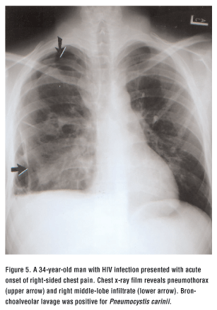
Pneumothorax as everyone know is the medical emergency especially when patient 's respiration is compromised.
Pneumothorax means air in the plural space.
Pneumothorax can be divided in primary or secondary causes.
Symptom for it is tachypnoeic,dyspnoeic and chest discomfort.
Primary pneumothorax usually happens in healthy patient without any lung pathology.
Usually happens in tall and thin patient.
secondary pneumothorax usually happens in patient with underlying pathology.
For example COAD(chronic obstrutive airway disease),cystic fibrosis,status asthmaticus
Infective lung disease such asPCP,necrotising pneumonia.interstitial lund disease such as sarcoidosis,tuberous sclerosis, idiopathic pul fibrosis.
connective tissue:Ankylosis spondylitis,polymyositis
Ca of lung,sarcoma
Treatment of choice is observation if pneumothorax is small
If it is big, insert chest tube
if small pneumothorax with severe resp distress, chest tube also need to be inserted.
pneumothorax resolve by 1.25% each day without intervention
Friday, July 28, 2006
contrast nephropathy
Everyone who got cronic renal failure presented with unstable angina or severe chest pain and require angioplasty intervention are warned about possible contrast nephropathy .
What the hell is that?
I will give you some brief account about that.
Cotrast nephropathy is impairment of renal function occured within 3 days of IV contrast.
Recovers in few days,Rarely irreversible.
Contrast nephropathy is peaked at 4-5 days and returns to baseline within 7-10 days. Renal function can persists up to 3 weeks.
Patient with impaired renal funtion.Iodinated contrast is nephrotoxic causing renal vasoconstiction and direct toxic effects upon renal tubules.
Is anyone with normal renal function carries the risk.
Risk factor for contrast nephropathy are
A:renal insufficiency
60% of pt develop contrast nephropathy had preexisting renal insufficiency.
The greater the impairment the greater the risk.
B:Diabetis mellitus(DM)
Pt with DM and preexisting renal insufficiency carries higher risk compare to nondiabetis with renal insufficiency develop contrart nephropathy.
But DM with normal renal function carries same risk as non diabetic pt.
C:Volume of contrast
The higher the amount the higher the risk
D:multiple myeloma
McCarthy found that is no increase incidence for nephropathy but previously they say yes.
How to prevent it
The main stay is hydration
Hydration with normal saline.
How about N acetyl cysteine?
Some evidence said benefit some say not.
What the hell is that?
I will give you some brief account about that.
Cotrast nephropathy is impairment of renal function occured within 3 days of IV contrast.
Recovers in few days,Rarely irreversible.
Contrast nephropathy is peaked at 4-5 days and returns to baseline within 7-10 days. Renal function can persists up to 3 weeks.
Patient with impaired renal funtion.Iodinated contrast is nephrotoxic causing renal vasoconstiction and direct toxic effects upon renal tubules.
Is anyone with normal renal function carries the risk.
Risk factor for contrast nephropathy are
A:renal insufficiency
60% of pt develop contrast nephropathy had preexisting renal insufficiency.
The greater the impairment the greater the risk.
B:Diabetis mellitus(DM)
Pt with DM and preexisting renal insufficiency carries higher risk compare to nondiabetis with renal insufficiency develop contrart nephropathy.
But DM with normal renal function carries same risk as non diabetic pt.
C:Volume of contrast
The higher the amount the higher the risk
D:multiple myeloma
McCarthy found that is no increase incidence for nephropathy but previously they say yes.
How to prevent it
The main stay is hydration
Hydration with normal saline.
How about N acetyl cysteine?
Some evidence said benefit some say not.
Subscribe to:
Posts (Atom)
